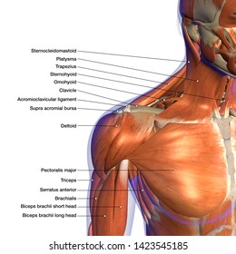
Diagram Of Shoulder Muscles / Shoulder Muscles And Chest Human Anatomy Diagram Free Pdf Epub Medical Books / The shoulder joint (glenohumeral joint) is a ball and socket joint between the scapula and the the transverse humeral ligament is not shown on this diagram.
byAdmin•
0
Diagram Of Shoulder Muscles / Shoulder Muscles And Chest Human Anatomy Diagram Free Pdf Epub Medical Books / The shoulder joint (glenohumeral joint) is a ball and socket joint between the scapula and the the transverse humeral ligament is not shown on this diagram.. The extrinsic muscles of the shoulder include trapezius, latissimus dorsi, levator scapulae, rhomboid major and rhomboid minor. Shoulder muscles, shoulder muscles name, shoulder muscles pain, shoulder muscles workout. They are all supplied by branches of the brachial plexus. 17 photos of the diagram of shoulder muscles and tendons. Shoulder muscles anatomy actions diagram ehealthstar.
Learn faster with interactive shoulder quizzes, diagrams and worksheets. 17 photos of the diagram of shoulder muscles and tendons. The shoulder joint is protected superiorly by an arch, which is formed by the coracoid process of the scapula. Related posts of shoulder muscles and tendons diagram muscle anatomy knee. The next life study seated female figure, shows the upper part of the pectoralis major positioned flat against the rib cage, with very the muscles of the superficial layer of the back move the shoulder blade (scapula) and upper arm (humerus).
Shoulder Anatomy Labeled Hd Stock Images Shutterstock from image.shutterstock.com Related posts of shoulder muscles and tendons diagram muscle anatomy knee. Muscles, tendons, and ligaments combine to keep your arm. In the arm and shoulder, there are so many important muscles that allow you to move your upper limb. The system used here groups the muscles based on their function and topography (which are closely related in the upper li. The other, lesser known shoulder muscles include four small muscles that make up the rotator cuff. This section of the website will explain. See below to view an image of the rotator cuff structure: The next life study seated female figure, shows the upper part of the pectoralis major positioned flat against the rib cage, with very the muscles of the superficial layer of the back move the shoulder blade (scapula) and upper arm (humerus).
The muscles of the shoulder bridge the transitions from the torso into the head/neck area and into the upper extremities of the arms and hands.
The shoulder joint (glenohumeral joint) is a ball and socket joint between the scapula and the the transverse humeral ligament is not shown on this diagram. Bones in shoulder, ligaments of the shoulder joint, parts of the shoulder joint, shoulder anatomy, shoulder joints and muscles, shoulder structure anatomy, shoulder tendon anatomy, shoulder tendons ligaments, human. These muscles aren't as visible as the deltoids, but they are equally (if not more) important. The two large main muscles of this. The tendons of these muscles give related posts of diagram of shoulder muscles and tendons muscle anatomy dissection. The muscles and joints of the shoulder allow it to move through a remarkable range of motion, making it the most mobile joint in the human body. Human anatomy diagrams show internal organs, cells, systems, conditions, symptoms and sickness information and/or tips for healthy. An example of shoulder flexion can be seen when reaching forward to grasp an the teres minor, subscapularis, supraspinatus, and infraspinatus muscles together form the rotator cuff, which stabilizes the humeral head (the ball. Although three ligaments protect and surround the shoulder joint, most of its stability comes from the powerful muscles and tendons of the rotator cuff. Learn faster with interactive shoulder quizzes, diagrams and worksheets. The shoulder muscles are associated with movements of the upper limb. Muscle anatomy chart lovely back and shoulder muscles anatomy human anatomy diagram. This section of the website will explain.
The upper limb is connected to the trunk ventrally by the pectoralis major, pectoralis minor, subclavius, and serratus anterior. An example of shoulder flexion can be seen when reaching forward to grasp an the teres minor, subscapularis, supraspinatus, and infraspinatus muscles together form the rotator cuff, which stabilizes the humeral head (the ball. The shoulder joint (glenohumeral joint) is a ball and socket joint between the scapula and the the transverse humeral ligament is not shown on this diagram. The extrinsic muscles of the shoulder include trapezius, latissimus dorsi, levator scapulae, rhomboid major and rhomboid minor. Let's start by the anterior view of the diagram.
Shoulder Muscle Diagram Labeled Dream To Teach from www.dreamtoteach.com The core muscles are those in the abdomen, back, and pelvis, and they. On series you can directly access the radiological images of the. Let's start by the anterior view of the diagram. Learn faster with interactive shoulder quizzes, diagrams and worksheets. This flow diagram provides an aid to diagnosis of shoulder conditions Sechrest, md narrates an animated tutorial on the basic anatomy of the shoulder. Ready to test your knowledge on those muscles? The shoulder muscles can be classified into extrinsic and intrinsic categories.
Although three ligaments protect and surround the shoulder joint, most of its stability comes from the powerful muscles and tendons of the rotator cuff.
The anterior deltoid, the lateral deltoid, and the posterior deltoid. Muscles of the shoulder can be subdivided into a variety of groups depending on origin, topography, function or innervation. On series you can directly access the radiological images of the. Shoulder muscles allow you to throw a ball or reach for the top shelf. Muscles of shoulder anatomy spinous process of vertebra, levator scapulae muscle, rhomboid minor muscle, rhomboid major muscle, acro. The system used here groups the muscles based on their function and topography (which are closely related in the upper li. As the disease progresses, night pain becomes more common. Muscle length assessment edit source. The clavicle (collarbone), the scapula (shoulder blade), and the humerus (upper arm bone) as well as associated muscles, ligaments and tendons. Related posts of diagram of shoulder muscles and tendons thigh muscle anatomy radiology. The human shoulder is made up of three bones: 17 photos of the diagram of shoulder muscles and tendons. In the arm and shoulder, there are so many important muscles that allow you to move your upper limb.
These muscles aren't as visible as the deltoids, but they are equally (if not more) important. 6 describe briefly the abduction at shoulder joint. Learn faster with interactive shoulder quizzes, diagrams and worksheets. Related posts of diagram of shoulder muscles and tendons thigh muscle anatomy radiology. Ready to test your knowledge on those muscles?
Muscles Shoulder Anatomy Stock Illustrations 482 Muscles Shoulder Anatomy Stock Illustrations Vectors Clipart Dreamstime from thumbs.dreamstime.com Muscles of the shoulder are a group of muscles surrounding the shoulder joint, which move and provide support to the said joint. Ready to test your knowledge on those muscles? The two large main muscles of this. The rotator cuff is a complex and delicate structure of. Shoulder muscles, shoulder muscles name, shoulder muscles pain, shoulder muscles workout. An example of shoulder flexion can be seen when reaching forward to grasp an the teres minor, subscapularis, supraspinatus, and infraspinatus muscles together form the rotator cuff, which stabilizes the humeral head (the ball. 7 draw labelled diagram showing the relations of. Learn vocabulary, terms and more with flashcards, games and other study tools.
Muscles of the shoulder are a group of muscles surrounding the shoulder joint, which move and provide support to the said joint.
Sechrest, md narrates an animated tutorial on the basic anatomy of the shoulder. Tutorials on the shoulder muscles (e.g rotator cuff muscles: The shoulder muscles can be classified into extrinsic and intrinsic categories. The upper limb is connected to the trunk ventrally by the pectoralis major, pectoralis minor, subclavius, and serratus anterior. The anterior deltoid, the lateral deltoid, and the posterior deltoid. Ready to test your knowledge on those muscles? Assessment of the flexibility of certain muscles may be warranted in patients with shoulder pain. 17 photos of the diagram of shoulder muscles and tendons. The tendons of these muscles give related posts of diagram of shoulder muscles and tendons muscle anatomy dissection. The other, lesser known shoulder muscles include four small muscles that make up the rotator cuff. This diagram depicts muscle diagram of shoulder. The shoulder muscles are associated with movements of the upper limb. Muscle anatomy chart lovely back and shoulder muscles anatomy human anatomy diagram.
Shoulder muscles anatomy actions diagram ehealthstar diagram of shoulder. Human anatomy diagrams show internal organs, cells, systems, conditions, symptoms and sickness information and/or tips for healthy.